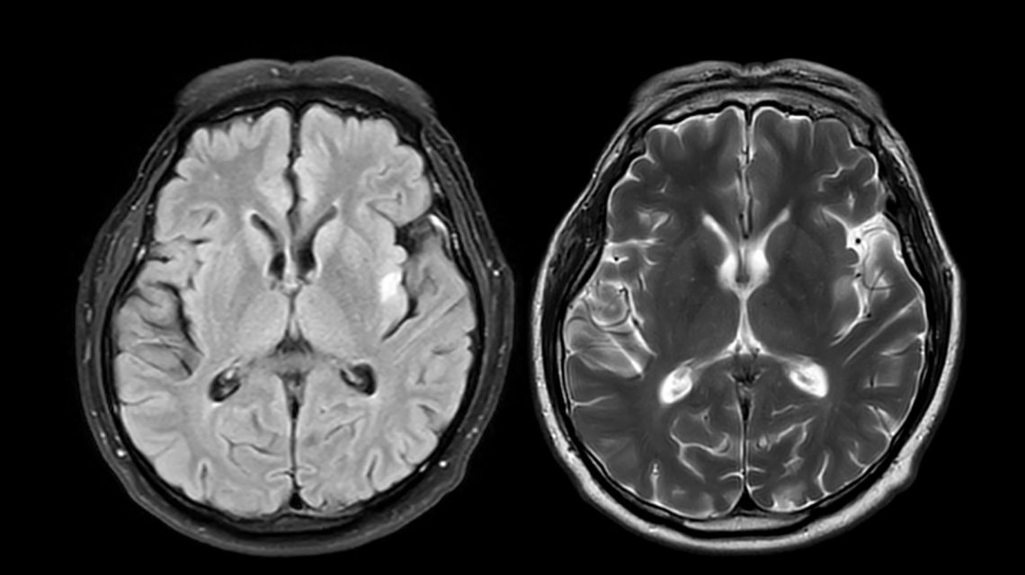
A new study suggests that a brain scan tool designed to measure shrinkage in areas linked to memory and thinking might be useful in predicting how quickly individuals with early-onset Alzheimer’s disease transition from mild cognitive impairment (MCI) to dementia. Early-onset Alzheimer’s disease typically manifests before age 65. It often begins with MCI, where memory and thinking issues are noticeable but not severe enough to be classified as dementia.
Researchers aimed to predict the speed of progression from MCI to dementia through patterns of brain shrinkage. To achieve this, they developed a tool called the early-onset Alzheimer’s disease signature, which detects atrophy in specific brain regions associated with thinking and memory, like parts of the parietal and temporal cortex.
The team measured the thickness of these areas using MRI scans and combined the results into a score reflecting shrinkage levels. The study cohort included 130 individuals with MCI living independently alongside 97 healthy participants, all aged 40 to 64. Each subject underwent an MRI scan at the start and attended annual follow-up visits, averaging two years in duration.
During the study, roughly 65% of participants with early-onset Alzheimer’s progressed to dementia. Increased gray matter shrinkage correlated with faster progression. An increase of one standard deviation in shrinkage raised the risk of transitioning from MCI to dementia by 1.24 times.
Notably, the brain scan biomarker predicted progression more accurately than just evaluating a participant’s symptoms initially. Lead researcher Alexandra Touroutoglou from Harvard Medical School noted that predicting the timeline of dementia progression remains challenging. The study results suggest the biomarker could function similarly to a timer, aiding doctors in estimating the onset and progression speed of dementia.
Dr. Ronald Schwartz from Continuum Health Services at Masonicare emphasized the potential value of this tool. He explained that while early-onset Alzheimer’s is known to be progressive, predicting individual progression rates is difficult. The tool might also aid patients and families in decision-making regarding clinical trials for treatments, work, finances, and future support.
However, Schwartz cautioned against viewing the prediction tool as a definitive answer. It should facilitate improved dialogue rather than serve as a precise forecast. He noted the biomarker maps brain regions exhibiting shrinkage in early-onset Alzheimer’s compared to those without the disease. This map can be applied to MRI scans to measure shrinkage and estimate progression.
Nonetheless, the researchers acknowledge that the biomarker was assessed within a single study group, predominantly non-Hispanic Whites, thus potentially limiting its applicability to diverse populations. Further research is necessary to ensure the tool’s accuracy across different demographics, disease stages, and clinical presentations.
The study advocates the potential of MRI-based gray matter shrinkage measurement in enhancing understanding of Alzheimer’s progression, despite needing broader validation. A more reliable tool may assist in setting expectations, planning care, and informing decisions for patients and families.
More information can be found in the study by Paranhos et al., published in Neurology: DOI: 10.1212/WNL.0000000000218519.
 Increased Funding Needed for Psychiatric Hospitals
  Trump Administration Distributes ACA Refund Checks
  Higher Consumption of Ultra-Processed Foods Linked to Increased Health Risks
  Understanding Inflammation: Myths and Facts
  Calm Parenting: Strategies to Manage Stress
  Trump’s Executive Order to Combat Mosquitoes and Ticks in D.C.